Home
/ Anatomy Of Musckes Sndctendons / Human Animal Anatomy And Physiology Diagrams Lower Back Anatomy Muscles Neck And Shoulder Muscles Muscle Anatomy Shoulder Anatomy - Anatomy, function, and rehab considerations.
Anatomy Of Musckes Sndctendons / Human Animal Anatomy And Physiology Diagrams Lower Back Anatomy Muscles Neck And Shoulder Muscles Muscle Anatomy Shoulder Anatomy - Anatomy, function, and rehab considerations.
Anatomy Of Musckes Sndctendons / Human Animal Anatomy And Physiology Diagrams Lower Back Anatomy Muscles Neck And Shoulder Muscles Muscle Anatomy Shoulder Anatomy - Anatomy, function, and rehab considerations.. The tendons of these muscles pass through a small corridor in the wrist known as the carpal tunnel. This is a table of skeletal muscles of the human anatomy. The muscles of the torso, examined in the previous chapter, include a few that attach directly into the upper arm and help move the humerus at the shoulder joint. From anterior to posterior, the tongue has 3 surfaces: Convergent muscles contain fibers that have a wide origin, but converge in order to attach to a narrow tendon.
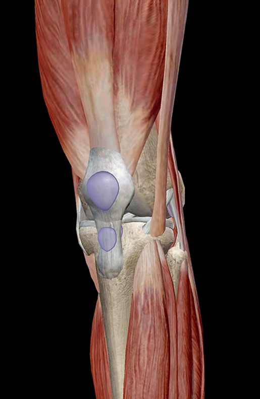
Anatomy of a muscle cell. Attached to the bones of the skeletal system are about 700 named muscles that make up roughly half of a person's body weight. You can click the links in the image, or the links below the image to find out more information on any muscle group. See the pictures and anatomy description of knee joint bones, cartilage, ligaments, muscle and tendons with resources for knee problems & injuries. The tongue is a mass of muscle that is almost completely covered by a mucous membrane.
Foot Anatomy Muscles Ankle Anatomy Ankle Tendonitis Foot Anatomy from i.pinimg.com In this section, learn more about the anatomy of the muscles of the neck. Skeletal muscles are attached to bones by tendons and can be as long as 30 cm, although they are usually 2 to 3 cm in length. Muscle tendons are extremely important in reinforcing and stabilizing joints. The anterior and middle scalenes originate from the transverse processes of certain cervical vertebrae and attach to the first rib. Find the best weight lifting exercises that target each muscle or groups of muscles. The muscular system consists of the skeletal muscles and their associated structures. Each of these muscles is a discrete organ constructed of skeletal muscle tissue, blood vessels, tendons, and nerves. As the skeletal muscles pull on bones to cause movements, they also stabilize the joints of the skeleton;
Smooth muscles (involuntary muscles) are usually in sheets or layers, with one layer of muscle behind the other.
This article will focus on tongue embryology, origin, insertion, and action of the extrinsic muscles, followed by innervation, blood supply and lymphatic drainage of the tongue. Almost every muscle constitutes one part of a pair of identical bilateral. General functions of muscular system: Inflammation of this region caused by repetitive stress or trauma may lead to pain and numbness known as carpal tunnel syndrome. Circular skeletal muscles are made up of fibers that are arranged in a circular manner. The three scalene muscles are found forming the floor of the posterior triangle. The tip is the highly mobile, pointed anterior portion of the tongue. See the pictures and anatomy description of knee joint bones, cartilage, ligaments, muscle and tendons with resources for knee problems & injuries. Skeletal muscles allow the body to move and maintain posture; In the muscular system, muscle tissue is categorized into three distinct types: Anatomy of the short head of the biceps brachii muscle. In this section, learn more about the anatomy of the muscles of the neck. Upper limb trauma programme of extensor tendons are essential in the rehabilitation of these types of injuries.
In the diagrams below, i'll be showing muscle groups in color, with a black line to show the forms that would show through the skin (i also show protruding bones that would do the same). Convergent muscles contain fibers that have a wide origin, but converge in order to attach to a narrow tendon. An interactive tutorial teaching the position, actions, innervation and attachments of the rectus femoris muscle with the aid of anatomical illustrations. Discover the muscle anatomy of every muscle group in the human body. Muscular contraction is necessary for voluntary and involuntary movement of limbs, stabilization of joints, maintaining luminal diameter (in the case of arteries, bowel, etc), and to produce heat.
Muscle Anatomy Of The Plantar Foot Everything You Need To Know Dr Nabil Ebraheim Youtube from i.ytimg.com Learn more about how muscles work, what they look like, and how they're treated. Anatomy of the muscular system. Muscle tendons are extremely important in reinforcing and stabilizing joints. The tendons of these muscles pass through a small corridor in the wrist known as the carpal tunnel. General functions of muscular system: By contracting, they also aid the venous return of blood to the heart and with age, these components of the musculoskeletal system progressively degenerate, which contributes to frailty and increases the risk of falls and fractures. This is a table of skeletal muscles of the human anatomy. Muscles of mastication are classified as main and accessory muscles.
General functions of muscular system:
Circular skeletal muscles are made up of fibers that are arranged in a circular manner. The tongue is a mass of muscle that is almost completely covered by a mucous membrane. Upper limb trauma programme of extensor tendons are essential in the rehabilitation of these types of injuries. Attached to the bones of the skeletal system are about 700 named muscles that make up roughly half. Muscle mass accounts for a large majority of the carcass weight of domestic animals. An interactive tutorial teaching the position, actions, innervation and attachments of the rectus femoris muscle with the aid of anatomical illustrations. Inflammation of this region caused by repetitive stress or trauma may lead to pain and numbness known as carpal tunnel syndrome. A collection of anatomy notes covering the key anatomy concepts that medical students need to learn. By contracting, they also aid the venous return of blood to the heart and with age, these components of the musculoskeletal system progressively degenerate, which contributes to frailty and increases the risk of falls and fractures. Learning to draw muscles may conjure medical charts in daunting details, but such complexity is unnecessary. Microscopic anatomy of skeletal muscle. Find the best weight lifting exercises that target each muscle or groups of muscles. As the skeletal muscles pull on bones to cause movements, they also stabilize the joints of the skeleton;
It elevates and protrudes the mandible. As the skeletal muscles pull on bones to cause movements, they also stabilize the joints of the skeleton; An interactive tutorial teaching the position, actions, innervation and attachments of the rectus femoris muscle with the aid of anatomical illustrations. It occupies most of the oral cavity and oropharynx. Anatomy of the muscular system.
Learn Muscle Anatomy Knee Joint Group from www.visiblebody.com There's no strict demarcation or dividing line between the tendon and the covering around this muscle but that covering is called is called the epimysium fp my cm and it's really just connective tissue that covers the muscle kind of protects it reduces friction. Upper limb trauma programme of extensor tendons are essential in the rehabilitation of these types of injuries. This is a table of skeletal muscles of the human anatomy. Microscopic anatomy of skeletal muscle. In the muscular system, muscle tissue is categorized into three distinct types: Adducts & flexes the arm (humerus). It elevates and protrudes the mandible. Attached to the bones of the skeletal system are about 700 named muscles that make up roughly half of a person's body weight.
Related online courses on physioplus.
The tip is the highly mobile, pointed anterior portion of the tongue. Smooth muscles are found in the walls of many organs, such as the stomach and in blood vessels. Discover the muscle anatomy of every muscle group in the human body. Skeletal muscles allow the body to move and maintain posture; Muscle tendons are extremely important in reinforcing and stabilizing joints. Related online courses on physioplus. Cardiac muscle contracts the heart to pump blood. Muscles of mastication are classified as main and accessory muscles. Convergent muscles contain fibers that have a wide origin, but converge in order to attach to a narrow tendon. An interactive tutorial teaching the position, actions, innervation and attachments of the rectus femoris muscle with the aid of anatomical illustrations. A collection of anatomy notes covering the key anatomy concepts that medical students need to learn. Microscopic anatomy of skeletal muscle. Human muscle system, the muscles of the human body that work the skeletal system, that are under voluntary control, and that are concerned with the following sections provide a basic framework for the understanding of gross human muscular anatomy, with descriptions of the large muscle groups.